Edited March 5, 2016 by Dr. Sudhir Trivedi, Director of R&D
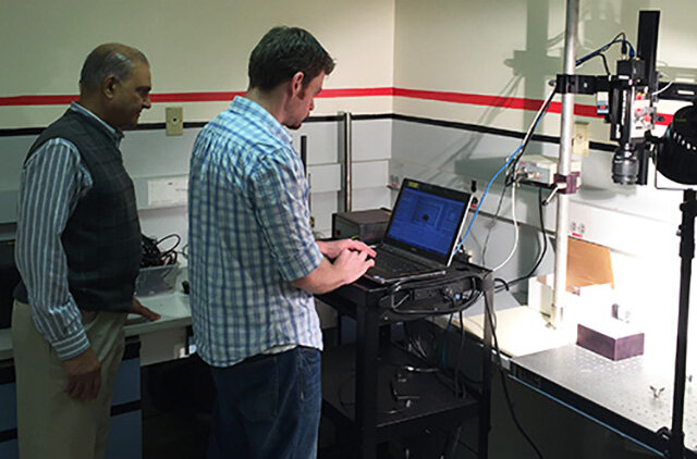
Brimrose Research Engineers evaluate a culture sample using an acousto-optic tunable filter hyperspectral imager.
Throughout the past 20 years or so, Brimrose has had a rich and storied history of providing its acousto-optic tunable filter (AOTF) technology to meet a variety of different needs, which can range from archeology to crime scene detection. Undoubtedly one of the most exciting applications is the detection of cancer, where our AOTF technology has been used in several ways.
In fact, the use of our AOTF technology in this area has given us a seat at the table of a fascinating new technology known as biomedical photonics, or BioPhotonics for short. This is changing the face of medicine, allowing us to view and operate in the human body as never before. More specifically, the use of our instruments is part of a new and growing field known as medical hyperspectral imaging, or MHSI. The use of MHSI does not require the introduction of agents, which is advantageous compared to imaging techniques that require contrast agents.
While I don’t want to get too technical, MHSI is a novel, camera-based method of imaging spectroscopy that integrates spatial and spectroscopic data from tissue in a set of images. MHSI delivers near-real-time images of biomarkers in tissue. It is a promising method for the noninvasive, rapid and inexpensive evaluation of cancer in the tumor bed.
A hyperspectral imaging system is mainly composed of the light source, wavelength dispersion devices and area detectors. The rationale for cancer detection by optical imaging lies in the fact that biochemical and morphological changes associated with lesions alter the absorption, scattering and fluorescence properties; therefore, optical characteristics of tissue can in turn provide valuable diagnostic information.
AOTF Technology: Improving Cancer Detection One Wavelength at a Time
Brimrose’s AOTF technology is a core technology for the company. AOTF technology allows us to scan hundreds of wavelengths per second, while also capturing critical data at any one or more of these precise wavelengths.
One of the first medical applications where our AOTF technology was used was for the Advanced Biomedical Science and Technology Group at Oak Ridge National Laboratory in Tennessee. The technology was adapted for the purpose of identifying skin cancer. This involved using the AOTF hyperspectral imager to detect cancerous tumors in mice.
The Oak Ridge researchers involved with the study were well pleased with the results: “Hyperspectral imaging sensors provide contiguous spectral signatures obtained from dozens or hundreds of narrow spectral channels. This enables hyperspectral imaging to reveal more useful information for material identification than conventional imaging techniques,” they found. (1)
Their conclusion: “The proposed hyperspectral fluorescence imaging technique can lead to the development of non-invasive in vivo detection of skin cancer without the need of tissue biopsy.” (2)
Another MHSI system with AOTF Brimrose provided to Chinese researchers was used in the evaluation of cancerous tongue tumors.
The results were rather extraordinary: A recognition rate of 96.5 percent was achieved by the researchers. They concluded: “The experimental results demonstrate that the system has great potential as an important imaging technology for medical imaging devices that provide additional diagnostic information regarding tissues under investigation. Although the final diagnostic decision remains the burden of physicians, the system supports physicians during decision making.” (3)
The system designed also included fiber probes for image signal collection, an endoscope, a laser excitation source, an endoscopic illuminator, an intensified charge-coupled device color camera for reflectance detection, a similar camera for fluorescence imaging, and a spectrometer for single-point spectroscopic measurements.
The Brimrose AOTF product also was used with a hyperspectral imager for the detection of cancer in skin, the breast, lungs and the tongue in collaboration with Columbia University Hospital.
Hyperspectral imaging sensors provide contiguous spectral signatures obtained from dozens or hundreds of narrow spectral channels. This enables hyperspectral imaging to reveal more useful information for material identification than conventional imaging techniques.
AOTF: Increasing our Understanding of Numerous Cancers
For example, the ability of AOTF technology used with hyperspectral imaging to provide a wavelength by wavelength analysis of tissue also sets it apart from multispectral imaging, which does not provide that degree of detail. This is critical as different types of cancer are found in focused, limited spectral ranges. For example, ovarian cancer is detected in the 400-640 nm spectral range, prostate cancer in the 450-950 nm range, melanoma in the 364-800 nm spectral range, etc. (4)
Medicine is slowly but methodically discovering the secrets of disease in the human body. An important way to do that is through ways to better understand what is going on in the human body through imaging processes. Hyperspectral imaging represents an important tool as we further make progress in discovering those secrets.
The future looks bright for MHSI. We expect that MHSI will play an important role for noninvasive disease diagnosis and monitoring, identification and quantitative analysis of cancer biomarkers, image guided minimum invasive surgery, targeted drug delivery and tracking and pharmaceutical drug dosage assessment.
Brimrose now offers the complete AOTF hyperspectral imaging unit, in both the SWIR and NIR ranges. Dr. Sudhir Trivedi is director of R&D at Brimrose Corporation, a position he has held for more than 20 years.
For more information about this blog, please call Brimrose at 410-472-7070 or send email to office@brimrose.com.
REFERENCES
“Hyperspectral Fluorescence Imaging for Mouse Skin Tumor Detection,” by Seong Kong, Matthew Martin and Tuan Vo-Dink, ETRI Journal, Volume 28, Number 6, p. 775.
Ibid
“Tongue Tumor Detection in Medical Hyperspectral Images,” by Zhi Liu, Hongjun Wang and Qingli Li, Sensors 2012, 12, 162-174.
“Medical Hyperspectral Imaging: A Review,” by Guolan Lu and Baowei Fei, Journal of Biomedical Optics, January 2014, 010901-4.